
Cranial Cruciate Injury in Dogs
The dog knee (stifle) is not all that different from a human knee. There are four ligaments primarily responsible for stability of the stifle – the cranial cruciate, caudal cruciate, medial collateral, and lateral collateral. The cranial cruciate ligament (CCL) is equivalent to the anterior cruciate ligament or Anterior Cruciate Ligament (ACL) in people.
What causes the CCL to tear?
Cranial cruciate ligament rupture is the most common orthopedic condition in dogs. Cranial cruciate ligament ruptures in dogs differ from ACL injuries in most people. Whereas trauma is a common cause of ACL tears in people, CCL tears in dogs is typically degenerative in nature. Young to middle-aged large female dog breeds are at the greatest risk of tearing their cranial cruciate ligament, though any dog can develop a CCL tear.
In most patients, once this degenerative process begins, the ligament will go on to a complete tear. Approximately half of all patients that go to surgery for repair of a CCL tear will still have a portion of the ligament left intact.
There are multiple hand-on tests your veterinarian can perform to help diagnose a cranial cruciate ligament tear. One of the first signs present prior to instability may be pain with full extension (hyperextension) of the knee. Once the ligament tears to a certain degree, the tibia can be manually manipulated to show instability in what is called the “cranial drawer test” or “tibial thrust” test in which the tibia can be moved forward in relation to the femur.
Other signs that may be noted in a physical exam include detection of effusion or swelling within the joint and scar tissue formation around the knee.
Though the cranial cruciate ligament is not visible on an x-ray, radiographs can help confirm a diagnosis of a tear by detection of changes that occur in the joint following this injury. These changes may include effusion (excess fluid in the stifle) and arthritis.
What are the treatment options for my dog?
As with many diseases, there are both medical and surgical treatment options for patients suffering from tears of the CCL. The choice to pursue surgical management is typically based on patient size, the stage of disease, the amount of instability present, the expectations you have for your pet’s activity level/quality of life. Surgery is the best long-term option.
Weight management and exercise modification are likely the most important things you can do to manage a patient with a CCL injury. Obesity has been found to quadruple the risk of CCL injury.
In large breed dogs, medical management has historically been thought to be overwhelmingly unsuccessful in returning them to full function. Surgical management consistently outperforms medical management and offers the most predictable long-term outcome.
What CCL surgery is best?
One option we offer at 2nd Street Animal Hospital is the traditional lateral extracapsular suture technique. This method may be recommended for small dogs if cost is an issue. This lateral extracapsular suture technique utilizes a nylon line to provide stability. Due to the low cost of the implant, the surgery is relatively inexpensive.
The 2nd option we offer is the Tibia Plateau Leveling Osteotomy (TPLO) technique. This involves changing the biomechanics of the knee joint via osteotomy or cutting of the bone. In essence, this osteotomy procedure alters the biomechanics of the joint and eliminates the need for the CCL. This surgery involves the use of a semi-circular bone cut at the top of the tibia and rotating it to decrease the Tibia Plateau Angle. The bone cut is then stabilized with a plate and screws, which are typically left in place unless there is a problem associated with the plate such as infection or irritation. This is a more expensive option and recommended for those dogs who are not a candidate for the first surgical option.
What are the risks of surgery?
As with any surgery, there are always potential complications. Anesthetic complications are possible, so it is important to consider a patient’s overall health prior to proceeding with surgery. Blood-work is recommended prior to surgery to ensure appropriate kidney and liver function, and to help screen for other diseases. Minor complications, such as swelling, bruising, or seroma formation are possible following surgery and typically self-limiting, able to be resolved within a few days of surgery.
Implant failure is extremely rare, but is the most catastrophic potential complication. Plates and screws used in canine TPLO are highly durable. However, in rare cases when pets are allowed too much activity very early on in the healing process, it’s possible for the plate and screws to bend or break.

Can this happen in the other leg?
About 50% of all dogs who suffer a ruptured cranial cruciate ligament in one knee will go on to rupture the same ligament in the opposite limb, typically within 1 to 2 years of rupturing the first side. Thankfully, the front legs of dogs are very similar to the human arm, so there are elbows, not knees, on the front limb.
What is recovery like for my dog?
Canine TPLO surgery involves making a bone cut (osteotomy), which is then stabilized with a bone plate and screws. In essence, we create a fracture and stabilize the bone in a new position. During this time we completely rely on the implants to stabilize the surgery site. Despite the invasiveness of this procedure, dogs are generally comfortable very quickly following surgery (24 to 48 hours) and begin weight bearing on the surgical limb within days. Though excessive activity can be destructive during the recovery period, frequent controlled weight bearing is invaluable to rapid bone healing.
The recovery period following a TPLO is approximately 8 weeks, as it takes approximately 8 weeks for bones to heal and good scar tissue to form.

Follow-Ups
Following surgery, a schedule will be set up for the first week for therapy laser treatment to reduce pain, inflammation and swelling.Your pet should return in 10 to 14 days from surgery to evaluate the healing of the incision. Sutures/staples were placed in the skin and will need to be removed during this time. If lameness occurs or your pet fails to improve, X-rays may need to be taken to verify proper implant position and evidence of healing. At 8 weeks post-surgery, X-rays will need to be taken to verify complete bone healing before returning to normal activity. The implants are designed to stay in for life. However, if pain, infection, or irritation occurs, we can easily remove them.
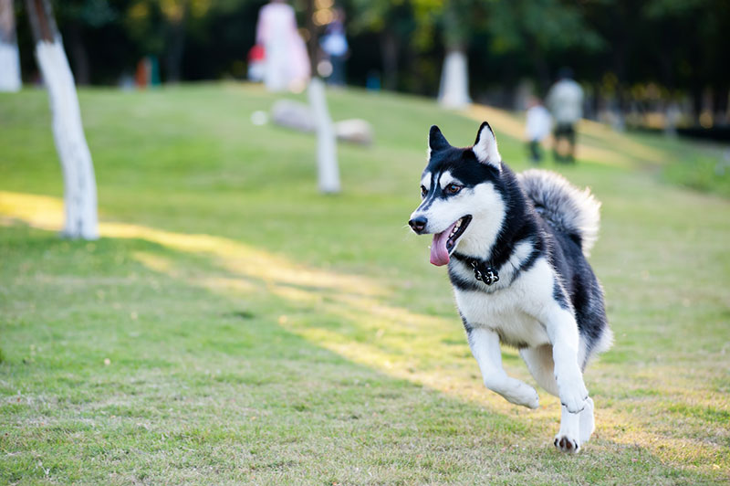
Will my dog ever run again?
There is a reported 90 to 95% chance of a good to excellent outcome with canine TPLO. This outcome indicates your pet will be able to run, jump, and play, and you will not notice your pet has ever had a problem. With a good outcome, after heavy activity your pet may have a short transient period of being sore and may need a short course of anti-inflammatories. For one reason or another, 5 to 10% of patients will not return to a level of function that we expect. It is important to note that just because your pet suffers a complication such as an infection or a meniscal injury, they can still have a good to excellent outcome. There just may be some hiccups along the way.